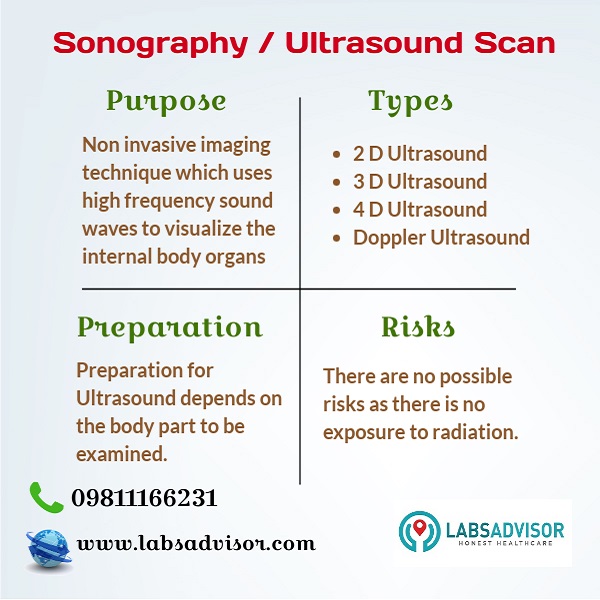
Sonography, known as Ultrasound Scan or USG is a diagnostic imaging technique that uses high-frequency sound waves to create images of the internal body organs such as kidneys, heart, abdomen, liver, and joints. It is a non-invasive medical test that helps doctors to detect the disease and treat medical conditions.
Book your Ultrasound scan in Ahmedabad at SRL Diagnostics, Surryam Imaging & at your local top quality labs through us at up to 50% discount. The lowest Sonography rate in Ahmedabad is ₹425 only.
Ultrasound Scan in Ahmedabad Through LabsAdvisor
|
Sonography Charges in Ahmedabad and Lab Details
Note that the sonography price in Ahmedabad mentioned may vary from the actual price. Click on the link to know the updated prices and lab details.
If you do not find your ultrasound scan in the above list, then call us at +918061970525. We will get back to you with the discounted Sonography price near you in Ahmedabad.
Get the lowest Sonography price in Ahmedabad by calling us at
To get a call back from our customer care team, click on the button below.

Frequently Asked Questions About Sonography / Ultrasound Scan
What is Sonography / Ultrasound Scan?
Sonography, known as Ultrasound Scan or USG is a diagnostic imaging technique that uses high-frequency sound waves to create images of the internal body organs such as kidneys, heart, abdomen, liver, and joints. It is a non-invasive medical test that helps doctors to detect the disease and treat medical conditions.
Unlike other radiology techniques, ultrasound has no exposure to radiation. Because of this, it is more commonly used to check the status of the fetus during pregnancy.
Different Types of Ultrasound Scan
The ultrasound scan can be broadly classified into the following four types on the basis of the type of image produced.
2D Ultrasound
2D Ultrasound is the most common type of sonography/ultrasound. It creates a series of 2-dimensional cross-section images of the tissues. The images produced are black and white and have the same kind of details as a photographic negative. The type of ultrasound is more often used throughout the pregnancy period to check the fetus and look for birth defects.
3D Ultrasound
3D ultrasound scans the tissue cross-section-wise at different angles and reconstructs the data received into 3D images. A common use of 3D ultrasound pictures is to closely examine the suspected fetal anomalies. 3D ultrasound results in a more effective diagnosis of abnormalities in the chromosomes.
4D Ultrasound
4D ultrasound pictures are created by updating 3D ultrasound images in quick succession. Due to the addition of time as the fourth dimension, the picture looks very realistic.
Doppler Ultrasound
Doppler ultrasound is a special type of ultrasound and a non-invasive test that uses high-frequency sound waves to estimate the blood flow through the blood vessels, especially the ones that supply blood to arms and legs. A regular ultrasound uses sound waves to produce images, but cannot estimate the blood flow.
It may be helpful to diagnose the following medical conditions
- Blood clots
- Defects in heart valves and congenital heart disease
- Bulging or narrowing of arteries
- Blockage in the artery
- Decreased blood circulation in legs
- Improper functioning of valves in the leg veins
Why is a Sonography done?
Your doctor may order an ultrasound or sonography if you experience pain, swelling, or other symptoms which require an internal view of the organs such as
- Bladder
- Brain in Infants
- Eyes
- Scrotum
- Kidneys
- Liver
- Ovaries
- Pancreas
- Spleen
- Thyroid
- Testicles
- Uterus
- Blood vessels
- Abdomen
- Pelvis
- Prostate
In addition to this, ultrasound is more commonly done at different stages of pregnancy to examine the health condition of the fetus. It is also very helpful to guide surgeons’ during certain medical procedures like biopsies.
Is there any preparation required before taking an Ultrasound?
The preparations for an Ultrasound scan vary depending on the study and the body part to be examined. In the case of abdominal ultrasound, you should fast for a minimum of 8 hours before the scan as the food particles may influence the accuracy of results.
In the case of bladder examination, the procedure requires a full bladder to get detailed information. So you should drink more water to fill the bladder and hold urine until the completion of the scan.
It is advised to consult your doctor regarding the preparations for your ultrasound study before the scan.
How is Sonography performed?
Sonography / Ultrasound is an imaging technique that uses high-frequency sound waves to visualize the internal organs. A device called a transducer is used to send sound waves into the body. Then the echoes are collected that come back after hitting the organs and sent to a special computer to process them into images.
The lab technician will ask you to change into a gown which will be provided in the lab. She will apply a lubricating gel on the body part to be examined to avoid any friction while rubbing the transducer against the skin. The images produced can be seen live on a computer screen.
Generally, an ultrasound scan takes up to 30 minutes to complete the entire procedure.4
Are there any risks in Sonography?
Sonography is a non-invasive technique done to detect abnormalities and examine the internal organs of the body. The sound waves which are sent into the body are harmless and there is no exposure to radiation like X rays. So it is a safe procedure to get a picture of the internal body parts. It is more commonly used in pregnancy to view the health condition of the unborn child.

How long does it take to get the Ultrasound reports?
The sonographer will analyze the images produced and prepare the reports. Usually, the ultrasound reports will be available within 30 minutes to 2 hours.
Interesting topics you may look for with LabsAdvisor:


