Sonography, also known as an ultrasound scan, is a diagnostic test that uses high-frequency sound waves to create an image of internal organs of the body such as kidneys, heart, liver, and joints.
Book this really important test at your local top-quality labs through us at up to 50% discount. The lowest Sonography test price in India is ₹200 only.
Sonography Scan in India Through LabsAdvisor
|
Sonography Test Price Near You and Lab Details
Below is the list of top Ultrasound scan prices in India, please click on the link of the Ultrasound scan you want and you can see all the labs near you and book online.
You can also select the time slot suitable for your USG scan. Click on the suitable link below to book anytime, 24 hours a day. Note that the prices mentioned below may vary from the actual.
If your Sonography scan is not listed in the table above, call us on +918061970525. We will get back to you with the Sonography charges near your location in India.
Get the lowest Sonography price in India by calling us at
If you want us to call you back, click on the link below
What is Sonography?
Sonography, also known as an ultrasound scan, is a diagnostic test that uses high-frequency sound waves to create an image of internal organs of the body such as kidneys, heart, liver, and joints. It is a non-invasive medical test that helps doctors to diagnose the disease and treat medical conditions.
Ionizing radiation (as used in x-rays) is not used in Ultrasound and therefore the radiation exposure to the patient is nil. The procedure is very safe and will not cause any pain.
Types of Sonography or Ultrasound Scan
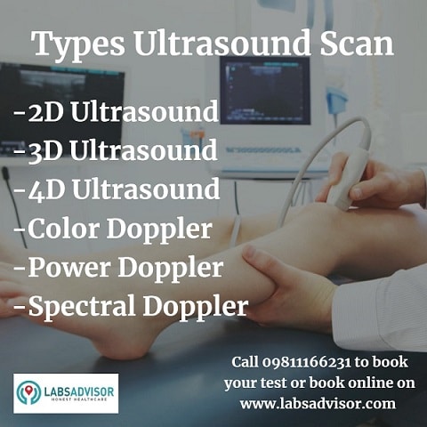
The ultrasound scan may be broadly classified into four types depending upon the type of ultrasound image. The choice of an Ultrasound scan depends upon the requirement of the investigation and availability of the equipment.
2D Ultrasound
2D Ultrasound is the most common type of ultrasound, it creates a series of flat 2-dimensional cross-section images of the tissue being scanned. The images from a 2D ultrasound are black and white and have the same kind of detail as a photographic negative. The 2D ultrasound is frequently used throughout pregnancy to check the fetus and look for birth defects.
3D Ultrasound
3D ultrasound is achieved by scanning tissue cross-sections at different angles and reconstructing the data received into three-dimensional images. A common use for 3D ultrasound pictures is to closely examine for suspected fetal anomalies. Other more subtle features such as low-set ears, cleft lip, deformation of face, or clubbing of feet can be better assessed, resulting in a more effective diagnosis of chromosomal abnormalities.
4D Ultrasound
Recently, 4-D or dynamic 3-D scanners are in the market. By updating 3D ultrasound images in quick succession, 4d ultrasound pictures are created. Due to the addition of time as the fourth dimension, the picture looks more realistic.

Doppler Ultrasound
In order to see how the blood flows through a blood vessel, doppler ultrasound emits high-frequency sound waves, usually the ones that supply blood to arms and legs.
When is a Doppler Ultrasound used?
- Blood clots
- Heart valve defects or congenital heart disease
- Bulging or narrowing of arteries
- A blocked artery
- Decreased blood circulation in legs
Doppler ultrasound may be done as an alternative to an invasive procedure such as arteriography or venography, which involves injecting dye into the blood vessels.
There are Three Types of Doppler Ultrasound:
Color Doppler – Uses a wide choice of color to visualize the blood flow measurement. It provides a pronounced representation of blood flow speed and direction as compared to the normal Doppler scan.
Power Doppler – It provides a more detailed representation of blood flow than the regular color Doppler. It can sometimes achieve images not accessible by color Doppler, however, color Doppler cannot indicate the direction of the blood flow.
Spectral Doppler – can determine the amount of blood flow direction and but it is represented in a graphical form instead of grayscale or color images.
Purpose of a Sonography or Ultrasound Scan
Ultrasound imaging assesses organ damage due to illness and helps diagnose various medical conditions. The imaging process is also helpful in guiding biopsies.
Doctors generally advise ultrasound to evaluate the following symptoms:
- Pain
- Swelling
- Infection
Uses of Ultrasound in different areas of medical science:
Ultrasound in an emergency – In case of an emergency, the damage caused to the organs is easily detected by ultrasound scan. It can help detect fluid build-up near the heart and blood in peritoneal cavities.
Gastroenterology – Ultrasound is used to detect appendicitis which is an inflammation of the appendix in the abdominal cavity. If detected, surgery to remove the appendix is advised. It is also used to see the images of other solid organs of the abdomen like kidneys, liver, pancreas, spleen, bile ducts, and inferior vena cava for any abnormalities.
Obstetric Sciences – Ultrasound is used to create an image of a fetus or embryo in the uterus. Obstetric sonography is a standard prenatal procedure. It is used to assess the progress of the pregnancy. Check the ultrasound scans for pregnancy to know the details of the scans for all the trimesters including the cost of an Ultrasound scan for pregnancy in India.
Urology – Ultrasound is used in urology also. Sometimes urine is left in the bladder after a person has urinated. People who cannot empty their bladder suffer from petty bladder muscular tone, or from other conditions that restrict the flow of urine, like bladder stones or an enlarged prostate. The Ultrasound is used to determine the amount of urine left in the bladder after urinating and then the diagnosis is done accordingly.
Organs in the pelvic region like the uterus can be checked. Pelvic sonography can be done internally or externally. In females, the sonogram may be inserted transvaginally and in males, it is inserted through the rectum. It can help the doctor determine the reason for incontinence or obstructed defecation.
Musculoskeletal Sonography – Ultrasound can be used to examine ligaments, soft tissues, nerves, muscles bone surfaces, and tendons. It is carried out to diagnose sprains, tears, strains, and the conditions of other soft tissue.
Neonatology – Ultrasound can be done in newborn infants to check for abnormalities in the brain and other body parts.
To book and know more about Ultrasound scan cost in India call +918061970525 or book an Ultrasound scan online.
Cardiology: Ultrasound is widely used in cardiology. An echocardiogram (ECG), also known as cardiac ultrasound, takes two-dimensional or three-dimensional images of the heart. The blood flow speed, the condition of cardiac valve areas, leaking of blood from the valves, and dis-functioning of right and left side of the heart. The overall working of the heart can be assessed through the echocardiogram.
Ultrasound for Treatment: Apart from diagnosis ultrasound is sometimes used to treat some illnesses.
- Dentists use Ultrasound to clean the teeth.
- Low-intensity ultrasound can stimulate bone growth.
- Ultrasound is helpful in breaking up stones in the kidney, ureter, or bladder with the help of its shock waves. The tiny pieces of stones are passed out of the body in urine.
- Cataracts can be treated with ultrasound waves.
- High-intensity focused ultrasound can be used to generate heat to treat tumors and cysts.
The Science Behind Sonography or Ultrasound Scan
Ultrasound imaging is done on the basis of the principle of reflection of ultrasonic waves. When a sound wave hits a thing, it returns back, and hence it is possible to determine the object size, shape, and consistency by measuring the echoed waves.
Ultrasound is helpful in detecting tumors and assessing the changes in appearance, size, and fluidity of tissues, organs, and blood vessels.

In the ultrasound procedure, the ultrasound gel and a small transducer (probe) are directly applied to the skin. High-frequency waves are transmitted from the transducer through the gel into the body. The sensitive microphone in the transducer records changes in sound pitch and direction as the sound waves bounce back from the organs, tissues, and fluids. This data is sent to the computer which creates real-time images and displays it on the monitor. One or more frames of the moving picture are captured and saved as scan reports.
How should I Prepare for the Procedure?
The preparation for the procedure depends on the type of scan. For some scans, your doctor may instruct you not to eat or drink as long as 12 hours before the appointment. For others, you may be asked to drink as many glasses of water in two hours before the scan and avoid urinating in order to keep your bladder full.
Wear loose-fitting clothes for your ultrasound scan.
How is it done?
Usually, for an ultrasound procedure, you will be asked to lie face up on the examination table. you may also be asked to turn to either side for better quality images. After you lay down, a water-based gel will be applied by your sonographer to the area to be examined. The gel helps the transducer make secure contact with your sin and eliminate air bubbles. The transducer is then placed on the body and moved back and forth until the desired image is captured.
You might feel a little pain from the transducer moving on a tender area, otherwise, the procedure causes no discomfort. After the imaging procedure, the gel is wiped off. The gel will not leave any stain on your clothes.
You may be asked to dress and wait while the report is reviewed and printed. You will get your report immediately so that you may resume your normal activities.
Benefits of Ultrasound Scan
The ultrasound scan has its long list of benefits
- Most ultrasounds are non-invasive. No poking of needles.
- Is a painless procedure.
- Is totally safe and not use any ionizing radiation. It is a highly preferred imaging technique for infants and pregnant ladies.
- Is easily available and less expensive than other imaging procedures
- It gives a clear picture of soft tissue in which X-rays are unable to show up.
- Because of its capability to give real-time imaging, it is used to guide procedures like needle biopsies and fluid aspiration.
Limitations of Ultrasound Scan
Ultrasound has difficulty penetrating bones and, therefore shows up only the outer surface of bone structures and not what lies within. To examine the internal structure of bones and joints MRI is typically used.
Obese patients are difficult to examine through ultrasound as a large amount of tissue weakens the sound waves.
Ultrasound cannot be used to study any part of the body containing air, so it is not a useful tool for diagnosing lung problems and those of the digestive tract. In such cases, barium extracts, CT scanning (check here to know more about CT Scan in India), and MRI (Check here to know more about MRI in India) are preferable.
Other topics you may be interested in:-


